torus palatinus ct
These growths are solid when you touch them and cannot be moved easily with your finger. This focal hyperostosis is covered by a thin and poorly vascularised layer of mucosa.

Torus Palatinus Radiology Case Radiopaedia Org
Mit zunehmendem Alter kann er jedoch an Größe zunehmen.

. Der Torus palatinus lat. The core of TP will appear as a dense and lobulated exostosis on radiographs and CT images Figures 45. Removal is required only if they are symptomatic.
These growths come in many different shapes and they may be very small or quite large. Es tritt am häufigsten bei Frauen und Frauen asiatischer Herkunft auf. The torus palatinus is a unilocular or multilocular exostosis that occurs in the midline of the hard palate.
Etwa 20 bis 30 Prozent der Bevölkerung haben Torus palatinus. Characteristics of Torus Palatinus The protrusion usually appears during early adulthood and gradually reduces in size as the bone gets re-absorbed with time. Both exostoses usually appear in the form of a series of swellings summary by Suzuki and Sakai 1960.
A torus palatinus TP is a bony protuberance located at the union of the processus palatinus maxillaes that form the hard palate. Fractures through tori have been reported 4. Torus palatinus TP is a spindle-shaped bony elevation along the midline of the hard palate.
2 article feature images from this case 3 public playlist include this case Promoted articles advertising. Torus Wulst palatum Gaumen ist eine Exostose knöcherner Längswulst entlang der Mittellinie des harten Gaumens. It is a bony proturberance almost always in the midline.
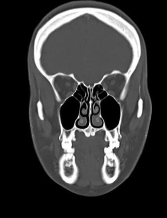
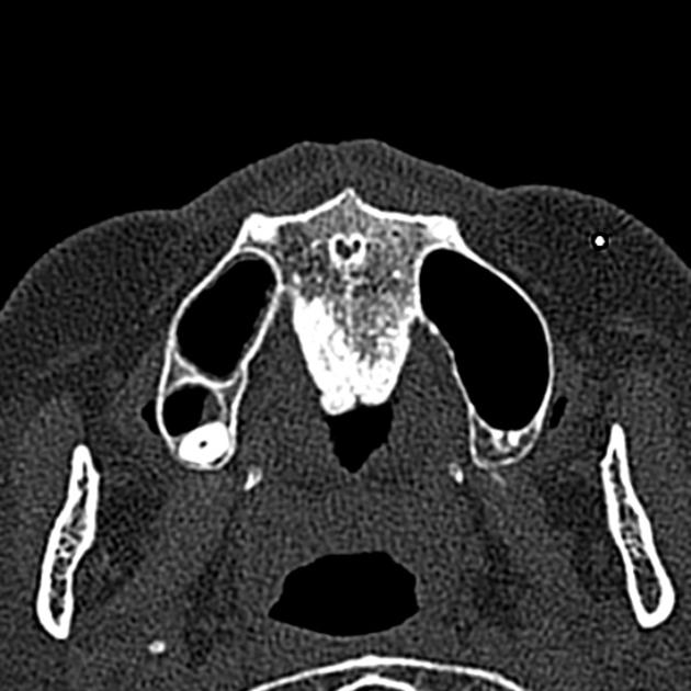
If imaged CT is the primary modality. Torus palatinus clearly appears as a bony protuberance arising from the inferior surface of the hard palate on coronal and sagittal CT images Fig. It is of no significance apart from patient concern usually about cancer and occasional denture rubbing leading to ulceration.
Typical appearance of hyperostosis in the midline hard palate either side of the suture. A torus palatinus is not cancerous. Treatment is usually conservative.
Treatment and prognosis Mandibular tori are benign slow growing and non-invasive. It is very important to know that torus palatinus and the use of BPs are risk factors for osteonecrosis of the maxilla. Magnetic resonance imaging MRI is not commonly used in dentistry and descriptions of the torus by this imaging method are therefore rare in the literature.
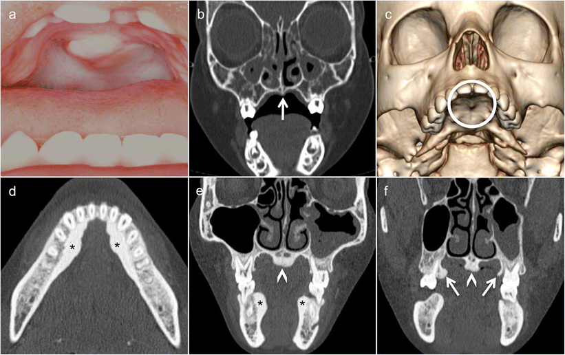
Torus mandibularis TM is a bony elevation along the mandible generally just below the lower premolars. Classifying torus palatinus according to their appearance is possible. Computed tomography CT was performed for confirmation of torus palatinus and to aid surgical planning.
What Is Torus Palatinus. The hard mass may be flat lobular nodular or spindle-shaped. See also mandibular tori.
Torus Palatinus Torus palatinus TP manifests as a fungating mass of variable size and configuration that protrudes into the oral cavity from the center of the hard palate. Torus palatinus ist ein harmloses schmerzloses Knochenwachstum das sich auf dem Gaumen dem harten Gaumen befindet. It is the palatine version of torus mandibularis.
FIGURE 1 OMITTED Discussion Torus palatinus is a benign reactive hyperplasia of osseous tissue extending. It is commoner in certain racial groups such as asians. A case report We point out the possible causative relationship of BPs and osteonecrosis on torus growth.
1 Definition Ein Torus palatinus ist eine Exostose des harten Gaumens. Die Masse erscheint in der Mitte des harten Gaumens und kann in Größe und Form variieren. Removal is required only if they are symptomatic.
8a while a lobulated appearance may be better observed on axial CT images Fig. It is considered a common clinical finding. Torus palatinus is largely a clinical diagnosis.
From the alveolar bone inner or outer from midline hard palate specifically from the palatine process of the maxilla inward protrusion into the oral cavity known as torus palatinus Treatment and prognosis Maxillary tori are benign slow growing and non-invasive. Different phenotypes of the TP can be recognised with a flat polylobulated spindle-shaped or nodular appearance. Radiographic features CT Bony outgrowths from the inner aspect of the alveolar bone of the mandible above the origin of the mylohyoid muscle.
A torus palatinus is a bony growth that develops on the roof of the mouth. The CT scan demonstrated lobulated bony outgrowths arising from the inferior margin of the hard palate consistent with torus palatinus. Torus palatinus are bony growths on the palate or roof of your mouth.
Inhaltsverzeichnis 1 Prothetik 2 Chirurgische Entfernung 3 Inzidenz 4 Einzelnachweise Prothetik Bearbeiten. 2 Hintergrund Beim Torus palatinus handelt sich um einen Knochenwulst im Bereich der Sutura palatina mediana der in der Regel einen Durchmesser von unter zwei Zentimeter hat. Torus palatinus osteonecrosis related to bisphosphonate.
The growths are non-cancerous and therefore not a threat to your life. 2 Medical reports show that torus palatinus affects more women than men. 1 article features images from this case Maxillary torus.

Cone Beam Ct Showing A Very Large Exophytic Torus Palatinus Arrows Download Scientific Diagram

Torus Palatinus Image Radiopaedia Org

Pdf An Incidental Radiographic Finding Torus Palatinus

Torus Palatinus Radiology Case Radiopaedia Org

Coronal A And Axial B Ct Scans Through The Palate Show A Torus Download Scientific Diagram

Torus Palatinus Radiology Case Radiopaedia Org

Torus Palatinus Radiology Case Radiopaedia Org

Torus Palatinus Radiology Case Radiopaedia Org

An Incidental Radiographic Finding Torus Palatinus

Pdf Imaging Aspects Of Palatal Torus In Cone Beam Computed Tomography And Magnetic Resonance Case Report Semantic Scholar

Progredient Wachsender Torus Mandibularis Zm Online
_359-364-f2.jpg)
Imaging Aspects Of Palatal Torus In Cone Beam Computed Tomography And Magnetic Resonance Case Report Abstract Europe Pmc
Bony Palate Keyword Search Science Photo Library

Ct Imaging Frontal View Prominent Exostosis Torus On The Hard Download Scientific Diagram

Masses Of Developmental And Genetic Origin Affecting The Paediatric Craniofacial Skeleton Insights Into Imaging Full Text


Comments
Post a Comment